文献速递:PET-影像组学专题--临床上在乳腺癌方面PET-MRI的进展
文献速递:PET-影像组学专题–临床上在乳腺癌方面PET-MRI的进展
01
文献速递介绍
成像在乳腺癌的筛查、诊断、分期和管理中扮演着关键角色。乳房X线摄影(mammography)、超声检查和动态增强乳腺MRI是主要的乳腺癌成像方法。对于怀疑或已知有转移性疾病的患者,使用CT、骨显像或PET-CT进行全身成像。PET-CT成像中用于乳腺癌患者的标准放射药物是2-[1?F]氟-2-脱氧-D-葡萄糖([1?F]FDG),它会在高糖代谢的组织中积聚。[1?F]FDG-PET-CT可用于初步分期新诊断的乳腺癌患者,以识别远处转移,重新分期怀疑复发或转移的患者,以及监测局部晚期和转移性乳腺癌患者的治疗反应。MRI也可用于对特定疾病部位(如大脑、脊柱或肝脏)的重点评估。长期以来,人们对结合PET与MRI进行更全面评估的潜在协同作用感兴趣。MRI提供高空间解剖细节、细胞密度和灌注数据,而PET提供功能性代谢信息。起初,通过将从俯卧位患者分别进行的动态增强乳腺MRI和PET-CT获得的图像进行共注册和融合,来结合这两种技术。随着2011年联合PET-MRI扫描仪的发展,现在可以在一次成像会话中完成。根据型号的不同,PET和MRI数据的获取可以顺序进行或同时进行。尽管使用了不同的衰减校正方法,但研究表明,通过PET-CT和同时进行的PET-MRI获得的放射药物摄取参数在患有原发性和转移性乳腺癌的患者中具有很强的相关性,验证了PET-MRI用于定量分析的有效性。在评估乳腺癌PET-MRI研究时,澄清成像协议中使用的获取类型很重要。全身PET-MRI是在患者仰卧位进行,使用表面射频线圈,而专用乳腺PET-MRI是在患者俯卧位进行,使用专用乳腺线圈(图1)。四通道、八通道和十六通道
使用了与集成PET-MRI扫描仪配套的四通道、八通道和十六通道射频乳腺线圈。成像协议还可以定制,以提供专门的腋窝PET-MRI检查,用于区域淋巴结分期。可以结合专用俯卧位乳腺PET-MRI扫描获得全身PET-MRI扫描,以评估局部、区域淋巴结和远处疾病。
PET-MRI在乳腺癌患者中提供附加价值的临床应用是一个活跃的研究领域。在本综述中,我们总结了有关PET-MRI在乳腺癌的诊断、分期、预后、肿瘤表型分析和治疗反应评估中的使用数据。
Title
题目
Clinical advances in PET–MRI for breast cancer
临床上在乳腺癌方面PET-MRI的进展
Conclusions
结论
PET–MRI has been studied for the diagnosis, staging,and tumour phenotyping of breast cancer, and for theassessment of prognosis and treatment response.
PET–MRI provides a comprehensive evaluation forpatients with newly diagnosed breast cancer, for whomthere is a clinical need to establish the extent of diseasein the breast and the locoregional lymph nodes and toestablish systemic staging at initial diagnosis orrecurrence. Importantly, PET–MRI has been shownto provide information that can change clinicalmanagement of the disease. Compared with PET–CT, thepatient is exposed to considerably less radiation fromPET–MRI, and this technique should, therefore, beconsidered, where available, for patients with clinicalindications for both PET–CT and organ-specific MRIexaminations. Based on the existing evidence, PET–MRIappears to be superior to PET–CT for the detection ofunsuspected extra-axillary nodal and distant disease,particularly hepatic and osseous metastases, in patientswith breast cancer.93 However, PET–MRI scanners arecurrently not as widely available as PET–CT scanners,which limits the accessibility of PET–MRI.94,95 Continuedresearch into targeted radiopharmaceuticals, radiomics,and methods to decrease radiation dose by reducing theactivity of injected radiopharmaceuticals will help toexpand the use of PET–MRI for breast cancer imaging inroutine clinical settings.
PET–MRI 已被研究用于乳腺癌的诊断、分期、
以及肿瘤表型鉴定,用于预后评估和治疗反应。
PET–MRI 为新诊断的乳腺癌患者提供全面评估,临床上需要确定乳腺及局部区域淋巴结的疾病范围,并在初次诊断或复发时建立全身分期。重要的是,PET–MRI 已被证明能提供可能改变疾病临床管理的信息。与 PET–CT 相比,患者接受的 PET–MRI 辐射量明显较少,
因此,在可行的情况下,对于临床上同时需要 PET–CT 和器官特异性 MRI 检查的患者,应考虑使用此技术。
根据现有证据,对于检测乳腺癌患者未预料的额外腋窝区域淋巴结和远处疾病,尤其是肝脏和骨转移,PET–MRI 似乎优于 PET–CT。93
然而,PET–MRI 扫描仪目前并不像 PET–CT 扫描仪那样广泛可用,这限制了 PET–MRI 的可及性。
94,95 对靶向影像性药物、影像组学,以及通过减少注射影像性药物的活性来降低辐射剂量的方法的持续研究,将有助于在常规临床环境中扩大乳腺癌成像的 PET–MRI 应用。
Figure
图

Figure 1: Simultaneous PET–MRI protocol
For dedicated breast PET–MRI with the patient in the prone position, PET data is acquired in one bed position during the MRI sequences (MRAC, T2, DWI, and T1 DCE).9 For whole-body PET–MRI with the patient in the supine position, PET data are acquired during MRI sequences in each of multiple (eg, six) bed positions.10 DCE=dynamic
contrast-enhanced. DWI=diffusion-weighted imaging. MRAC=magnetic-resonance-based attenuation correction. T1=T1-weighted imaging. T2=fluid-sensitive. T2-weighted imaging
图1:同步PET–MRI方案
对于患者采用俯卧位进行的专用乳腺PET-MRI,PET数据在MRI序列(MRAC、T2、DWI和T1 DCE)的一个床位位置上获取。
对于患者采用仰卧位进行的全身PET-MRI,PET数据在每个多个(例如,六个)床位位置的MRI序列期间获得。10 DCE=动态增强。DWI=弥散加权成像。MRAC=基于磁共振的衰减校正。
T1=T1加权成像。T2=对液体敏感的T2加权成像。

Figure 2: Dedicated prone-position breast 2-[1?F]fluoro-2-deoxy-D-glucose ([1?F]FDG)-PET–MRI showingcT2cN1 clinical anatomical staging Staging according to conventional techniques (ie, mammography, ultrasonography, and palpation) was cT2cN0.(A) Right breast invasive ductal carcinoma (maximum SUV=15·1). (B) Right level I axillary lymph node(maximumSUV=3·0).PathologicalstagepT2pN1a.SUV=standardised uptake value.
图 2:专用俯卧位乳腺 2-[1?F]氟代-2-脱氧-D-葡萄糖 ([1?F]FDG)-PET–MRI 显示cT2cN1 临床解剖分期 根据常规技术(即,乳房 X 光摄影术、超声检查和触诊)的分期为 cT2cN0。(A) 右侧乳腺浸润性导管癌(最大标准化摄取值(SUV)=15.1)。(B) 右侧 I 级腋窝淋巴结(最大SUV=3.0)。病理分期 pT2pN1a。SUV=标准化摄取值。

Figure 3: Dedicated prone-position breast 2-[1?F]fluoro-2-deoxy-D-glucose ([1?F]FDG)-PET–MRI showingcT3cN3b clinical anatomical stagingStaging according to conventional techniques (ie, mammography, ultrasonography, and palpation) was identifiedas cT2cN1. (A) Left internal mammary lymph node (maximum SUV=6·9). (B) Level I axillary lymph node(maximum SUV=14·4). ? Left breast invasive ductal carcinoma (maximum SUV=12·2). Pathological stage ypT3ypN2a. SUV=standardised uptake value
图 3:专用俯卧位乳腺 2-[1?F]氟代-2-脱氧-D-葡萄糖 ([1?F]FDG)-PET–MRI 显示 cT3cN3b 临床解剖分期 根据常规技术(即,乳房 X 光摄影术、超声检查和触诊)识别的分期为 cT2cN1。(A) 左侧内乳腺淋巴结(最大标准化摄取值(SUV)=6.9)。(B) I 级腋窝淋巴结 (最大SUV=14.4)。? 左乳浸润性导管癌(最大SUV=12.2)。病理分期 ypT3ypN2a。SUV=标准化摄取值。
Table
表

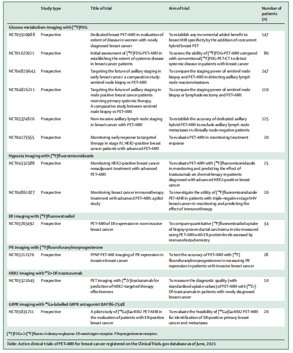
Table: Active clinical trials of PET–MRI for breast cancer registered on the ClinicalTrials.gov database as of June, 2021
inical trials of PET–MRI for breast cancer registered on the ClinicalTrials.gov database as of June, 2021*
表格:截至 2021 年 6 月,在 ClinicalTrials.gov 数据库上注册的用于乳腺癌的 PET–MRI 活跃临床试验
本文来自互联网用户投稿,该文观点仅代表作者本人,不代表本站立场。本站仅提供信息存储空间服务,不拥有所有权,不承担相关法律责任。 如若内容造成侵权/违法违规/事实不符,请联系我的编程经验分享网邮箱:veading@qq.com进行投诉反馈,一经查实,立即删除!