MITO-ID?线粒体膜电位检测试剂盒
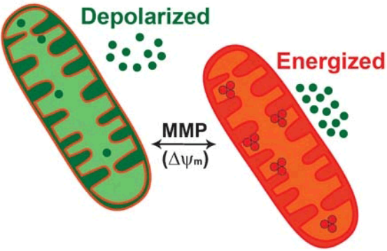
线粒体膜电位(Mitochondrial Membrane Potential,MMP)是判定细胞健康程度、线粒体膜通透性和细胞凋亡的一个重要指标,MMP的丧失通常与细胞凋亡的早期阶段有关。评估线粒体功能状态的基于细胞的检测方法正在成为阐明线粒体活动在药物诱导毒性、细胞凋亡级联以及其他细胞和生化过程中的作用的有用工具。
Enzo Life Sciences的MITO-ID??Membrane potential detection kit包含一种双发射阳离子染料,用于检测活细胞中的线粒体膜电位(MMP)。在有能量或活跃的细胞中,MITO-ID??膜电位试剂因其相对负电荷而在线粒体中迅速聚集成发橙色荧光的聚合体,而在细胞质中则以发绿色荧光的单体存在。然而,在MMP受损的细胞中,MITO-ID?膜电位试剂主要以绿色荧光单体存在于整个细胞质中,在线粒体中不再表现出橙色荧光。
作用机制
该染料的基本化学结构由高度共轭的部分组成,使正电荷广泛离域。该染料能够选择性地进入线粒体,当膜电位增加时,它的颜色会从绿色可逆地变为橙色(双发射电位探针)。这种光物理特性是由于在膜极化时可逆地形成J-聚集体,导致在490nm处激发时,发射光从~530nm转移到590nm。因此,具有低膜电位的线粒体将积累低浓度的染料,并表现出绿色荧光,而更高极化的线粒体将表现出橙色的荧光。
?MITO-ID??Membrane potential detection kit线粒体膜电位检测试剂盒
产品特点
●?耐光双发射染料,能够根据线粒体膜电位状态发出绿色或橙色荧光
●?比JC-1荧光染料灵敏10倍,并具有卓越的水溶性
●?无需洗涤步骤
●?适用于化学/环境毒性筛选
●?适用于高通量应用
实验示例
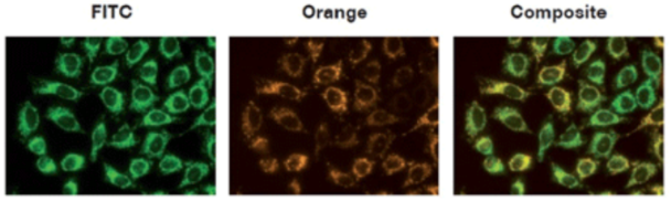
图1.?用MITO-ID??Membrane Potential reagent对HeLa细胞的线粒体进行染色,并通过荧光显微镜进行观察。橙色荧光聚集体定位于线粒体(橙色通道),而绿色荧光单体主要定位于细胞质(FITC通道)。
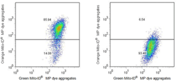
图2.?对照组和实验组细胞的流式细胞仪分析。未经处理的Jurkat细胞(左)与用1μM CCCP处理15分钟的Juekat细胞(右),用MITO-ID??Membrane Potential reagent对细胞进行染色,并使用流式细胞仪检测。
产品信息
| 产品货号 | ENZ-51018-0025/ ENZ-51018-K100 |
| 产品名称 | MITO-ID??Membrane potential detection kit? / 欣博盛生物 |
| 规格 | 1*25tests/1*100tests |
| 短期保存 | -20°C |
| 长期保存 | -80°C |
| 试剂盒组分 | MITO-ID??MP Detection Reagent Necrosis Detection Reagent CCCP Control 10X Assay Buffer 1 50X Assay Buffer 2 |
| 应用 | Flow Cytometry, Fluorescence microscopy, Fluorescent detection, HTS |
部分产品引用文献
1. Discovery of the 3-Amino-1,2,4-triazine-Based Library as Selective PDK1 Inhibitors with Therapeutic Potential in Highly Aggressive Pancreatic Ductal Adenocarcinoma: D. Carbone, et al.; Int. J. Mol. Sci.?24, 3679 (2023)?
2. Gene signature predicting recurrence in oral squamous cell carcinoma is characterized by increased oxidative phosphorylation: J.K. Noh, et al.; Mol. Oncol.?17, 134 (2023)?
3. Cu(I) and Cu(II) Complexes Based on Lonidamine-Conjugated Ligands Designed to Promote Synergistic Antitumor Effects: F.D. Bello, et al.; Inorg. Chem.?61, 4919 (2022)?
4. Mitochondrial membrane potential-enriched CHO host: a novel and powerful tool for improving biomanufacturing capability: L. Chakrabarti, et al.; MAbs?14, 2020081 (2022)
5. New glycoconjugation strategies for Ruthenium(II) arene complexes via phosphane ligands and assessment of their antiproliferative activity: D. Iacopini, et al.; Bioorg. Chem.?126, 105901 (2022)
6. New tricyclic systems as photosensitizers towards triple negative breast cancer cell: M. Barreca, et al.; Arch. Pharm. Res.?45, 806 (2022)?
7. Role of caspase-8 and/or-9 as biomarkers that can distinguish the potential to cause toxic-and immune related-adverse event, for the progress of acetaminophen-induced liver injury: T. Noda, et al.; Life Sci.?294, 120351 (2022),?Application(s):?Microplate reader?
8. Unveiling the Potential of Innovative Gold(I) and Silver(I) Selenourea Complexes as Anticancer Agents Targeting TrxR and Cellular Redox Homeostasis: M.D. Franco, et al.; Chemistry?28, e202201898 (2022)?
9. Altered inflammatory response in FMRP-deficient microglia: J.M. Parrott, et al.; iScience?24, 103293 (2021),?Application(s):?Fluorescence microscopy?
10. ApoE4 impairs neuron-astrocyte coupling of fatty acid metabolism: G. Qi, et al.; Cell. Rep.?34, 108572 (2021),?Application(s):?Microplate reader?
11. Botrytis cinerea methyl isocitrate lyase mediates oxidative stress tolerance and programmed cell death by modulating cellular succinate levels: L. Oren-Young, et al.; Fungal Genet. Biol.?146, 103484 (2021),?Application(s):?Conidia (fungus spore); microscopy?
12. Copper (II) complexes containing natural flavonoid pomiferin show considerable in vitro cytotoxicity and anti-inflammatory effects: J. Vanco, et al.; Int. J. Mol. Sci.?22, 7626 (2021),?Application(s):?Flow cytometry
13. Depletion of mitochondrial components from extracellular vesicles secreted from astrocytes in a mouse model of fragile X syndrome: B.G. Ha, et al.; Int. J. Mol. Sci.?22, 410 (2021),?Application(s):?Fluorescence microscopy
14. Inhibiting autophagy targets human leukemic stem cells and hypoxic AML blasts by disrupting mitochondrial homeostasis: K.M. Dykstra, et al.; Blood Adv.?5, 2087 (2021)
15. Selective striatal cell loss is ameliorated by regulated autophagy of the cortex: K. Cho & G.W. Kim; Life Sci.?282, 119822 (2021),?Application(s):?Flow cytometry
MITO-ID??Membrane potential cytotoxicity kit线粒体膜电位细胞毒性检测试剂盒
Enzo Life Sciences的MITO-ID??Membrane potential cytotoxicity kit利用阳离子双发射染料检测线粒体膜电位(MMP)的波动,该染料在细胞质中以绿色荧光单体的形式存在,在线粒体中以橙色荧光J聚集体的形式聚集。具有低膜电位的线粒体将积累低浓度的染料并呈现绿色荧光,而更高度极化的线粒体将呈现橙红色荧光。随着线粒体功能受损加剧,细胞表现出从橙色荧光到绿色荧光的转变。该试剂盒是一种独特的HTS测定法,无需洗涤或去除培养基即可实时监测线粒体膜电位。
产品特点
●?灵敏度是JC-1荧光染料的10倍,并具有卓越的水溶性
●?具备光稳定性的双发射染料
●?无需漂洗/换液步骤
●?单独的MITO-ID?检测可用于检测线粒体质量
●?可在较低的药物/剂量浓度下检测细胞毒性
●?没有使用JC-1染料时出现的溶剂伪影
●?适用于高通量应用
实验示例

图1.?检测线粒体紊乱的灵敏度是JC-1的10倍。使用MITO-ID?膜电位染料(红色)或JC-1(蓝色)在经CCCP处理的HeLa细胞中评估线粒体膜电位(MMP)。使用传统的荧光酶标仪检测,结果显示MMP?随着CCCP浓度的增加而减少,橙色荧光减少。染料的水溶性优化和无需洗涤的实验方案最大限度减小了可变性,因此Z因子(>0.9)高于使用JC-1.
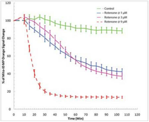
图2.?在药物筛选中实时检测有丝分裂毒性。使用BioTek Synergy? Mx荧光酶标仪对线粒体膜电位变化的时间进程研究。HeLa细胞与MITO-ID?膜电位染料在室温下孵育30分钟(不去除血清或培养基)。添加鱼藤酮分别达到1μM、3μM和9μM的浓度。橙色信号的减少证明染料对鱼藤酮有反应。
产品信息
| 产品货号 | ENZ-51019-KP002 |
| 产品名称 | MITO-ID??Membrane potential cytotoxicity kit / 欣博盛生物 |
| 规格 | 1 Kit |
| 短期保存 | -20°C |
| 长期保存 | -80°C |
| 试剂盒组分 | MITO-ID??MP Detection Reagent, 200 μL CCCP Control, 100 μL 10X Assay Buffer 1: 2.5 mL 50X Assay Buffer 2: 0.5 mL |
| 应用 | HTS |
??
部分产品引用文献
1. Novel Silver Complexes Based on Phosphanes and Ester Derivatives of Bis(pyrazol-1-yl)acetate Ligands Targeting TrxR: New Promising Chemotherapeutic Tools Relevant to SCLC Management: M. Pellei, et al.; Int. J. Mol. Sci.?24, 4091 (2023)?
2. FUNDC1 regulates receptor-mediated mitophagy independently of the PINK1/Parkin-dependent pathway in rotenone-treated SH-SY5Y cells: S.Y. Park, et al.; Food Chem. Toxicol.?137, 111163 (2020),?Application(s):?Fluorescence microscopy and microplate reader
3. Inhibitory role of TRIP-Br1/XIAP in necroptosis under nutrient/serum starvation: Z. Sandag, et al.; Mol. Cells?43, 236 (2020)?
4. NAD hydrolysis by the tuberculosis necrotizing toxin induces lethal oxidative stress in macrophages: D. Pajuelo, et al.; Cell. Microbiol.?22, e13115 (2020),?Application(s):?THP-1 macrophages; microplate reader
5. Impaired autophagic and mitochondrial functions are partially restored by ERT in Gaucher and Fabry diseases: M.M. Ivanova, et al.; PLoS One?14, e0210617 (2019),?Application(s):?Fluorescence microscopy?
6. Inhibitory role of AMPactivated protein kinase in necroptosis of HCT116 colon cancer cells with p53 null mutation under nutrient starvation: D.T. Le, et al.; Int. J. Oncol.?54, 702 (2019)?
7. ROS as a novel indicator to predict anticancer drug efficacy: T. Zaidieh, et al.; BMC Cancer?19, 1224 (2019)?
8. TREM1/3 deficiency impairs tissue repair after acute kidney injury and mitochondrial metabolic flexibility in tubular epithelial cells: A. Tammaro, et al.; Front. Immunol.?10, 1469 (2019),?Application(s):?Microplate reader
9. Anti-cancerous effect of cis-khellactone from Angelica amurensis through the induction of three programmed cell deaths: S. Jung, et al.; Oncotarget?9, 16744 (2018),?Application(s):?Microplate reader?
10. Blue light phototoxicity toward human corneal and conjunctival epithelial cells in basal and hyperosmolar conditions: V. Marek, et al.; Free Radic. Biol. Med.?126, 27 (2018),?Application(s):?Microplate reader
11. cGAS drives noncanonical-inflammasome activation in age-related macular degeneration: N. Kerur, et al.; Nat. Med.?24, 50 (2018)
12. Cytotoxicity of propofol in human induced pluripotent stem cell-derived cardiomyocytes: K. Kido, et al.; J. Anesth.?32, 120 (2018),?Application(s):?Microplate reader
13. Development of novel amino-quinoline-5, 8-dione derivatives as NAD (P) H: quinone oxidoreductase 1 (NQO1) inhibitors with potent antiproliferative activities: Y. Ling, et al.; Eur. J. Med. Chem.?154, 199 (2018)
14. Inhibition of apoptosis using exosomes in Chinese hamster ovary cell culture: S. Han, et al.; Biotechnol. Bioeng.?115, 1331 (2018)
15. Light action spectrum on oxidative stress and mitochondrial damage in A2E-loaded retinal pigment epithelium cells: M. Marie, et al.; Cell Death Disc.?9, 287 (2018)
本文来自互联网用户投稿,该文观点仅代表作者本人,不代表本站立场。本站仅提供信息存储空间服务,不拥有所有权,不承担相关法律责任。 如若内容造成侵权/违法违规/事实不符,请联系我的编程经验分享网邮箱:veading@qq.com进行投诉反馈,一经查实,立即删除!